 Impedance Medical Technologies
Electrical impedance mammography is a non-invasive, safe method of mammary glands examination. The examination is carried out with a multi-frequency electrical impedance mammograph MEM.
Electrical impedance mammography makes it possible to:
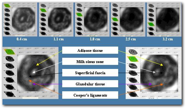
Figure 1. Electrical impedance anatomy of the mammary gland (50 kHz).
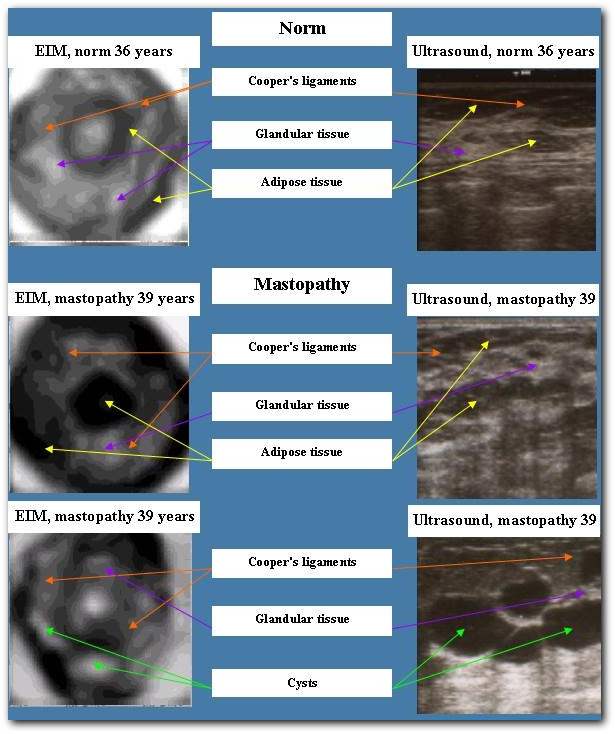
Figure 2. Electrical impedance mammography and ultrasound examination in the norm and in case of mastopathy.
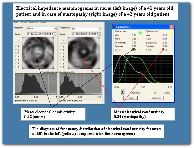
Figure 3. Indices of mean electrical conductivity in the norm and in case of mastopathy.
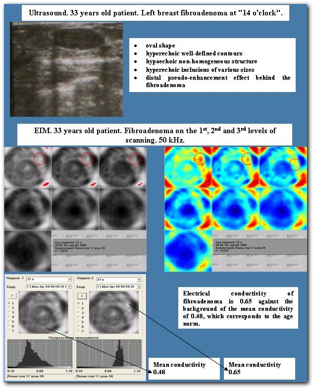
Figure 4. Mammary fibroadenoma.
Diagnose breast cancer (figures 5, 6)
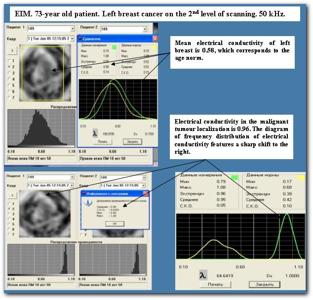
Figure 6. Left breast cancer