+7 (4852) 58-13-23
+7 (4852) 91-37-77
+7 (4852) 91-37-77
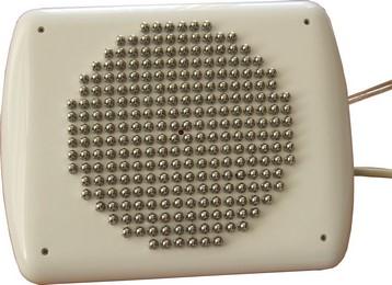
The device is intended for imaging and diagnostics of pathological changes of the breast tissues.
The electrical impedance method of diagnostics is based on the fact that electrical conductivity (ability to conduct electrical current) of biological tissues correlates considerably to their physiological status. It is well-known that many tumours, in particular malignant breast tumours, feature electrical conductivity, which differs significantly from the electrical conductivity of surrounding healthy tissues.
This phenomenon explains high sensitivity of the mammograph. The device enables, using the Electrical Impedance Tomography (EIT) method, to obtain the picture of biological tissues electrical conductivity distribution in the cros-sections of the breast at different frequencies. Distribution of electrical conductivity in every cross-section is visualized on a PC screen. The key advantages of electrical impedance method of diagnostic are as follows: absolute safety of examination, high level of information content, compactness, affordability and simplicity of the examination procedure.
The mammograph is intended for usage in conditions of specialized branches of hospitals and clinics. The device software supports exchange of data and images in DICOM format.
Various organs and tissues in a human body possess different electrical properties. For instance, it is a well-known fact that many tumours, in particular malignant breast tumours, feature electrical conductivity (ability to conduct electrical current), which differs considerably from the electrical conductivity of surrounding healthy tissues. The MEM makes it possible to obtain the picture of biological tissues electrical conductivity distribution in the breast in its transverse sections and locate such tumours in the obtained images.
.jpg)
.jpg)
.jpg)


The multifrequency electrical impedance mammograph (MEM) principle of operation is based on the method of electrical impedance tomography (EIT). The present device has come to replace the previously developed single-frequency device MEIK. Scientific research in the field of electrical impedance tomography has been developing since the middle of 1980s. The method enables the examiner, utilizing a full (in mathematical sense) set of electrical measurements, performed with the help of a multielectrode system, to reconstruct spatial distribution of electrical properties inside an object. The process of reconstruction is carried out through solving of the so-called inverse problem for the equation of electric field in inhomogeneous medium. A group of Russian scientists, comprising researchers from the Institute of Radio-engineering and Electronics of the Russian Academy of Sciences, is one of the world leader in the field of the EIT.
Below comes a list of some publications of the abovementioned researchers in scientific journals:


